Diagram Of Shoulder : Shoulder Bone Anatomy / If you have new, worsening, or severe shoulder pain, you should seek medical attention.. Inability to carry objects or use your arm. The most flexible joint in the entire human body, our shoulder joint is formed by the union of the humerus, the scapula (or shoulder blade), and the clavicle (or collarbone). The supraspinatus is located on the upper part of the shoulder joint and is involved in abduction (arm raising). Injuries to the rotator cuff are common, but treatment is often successful. Inside the shoulder there are three joints;
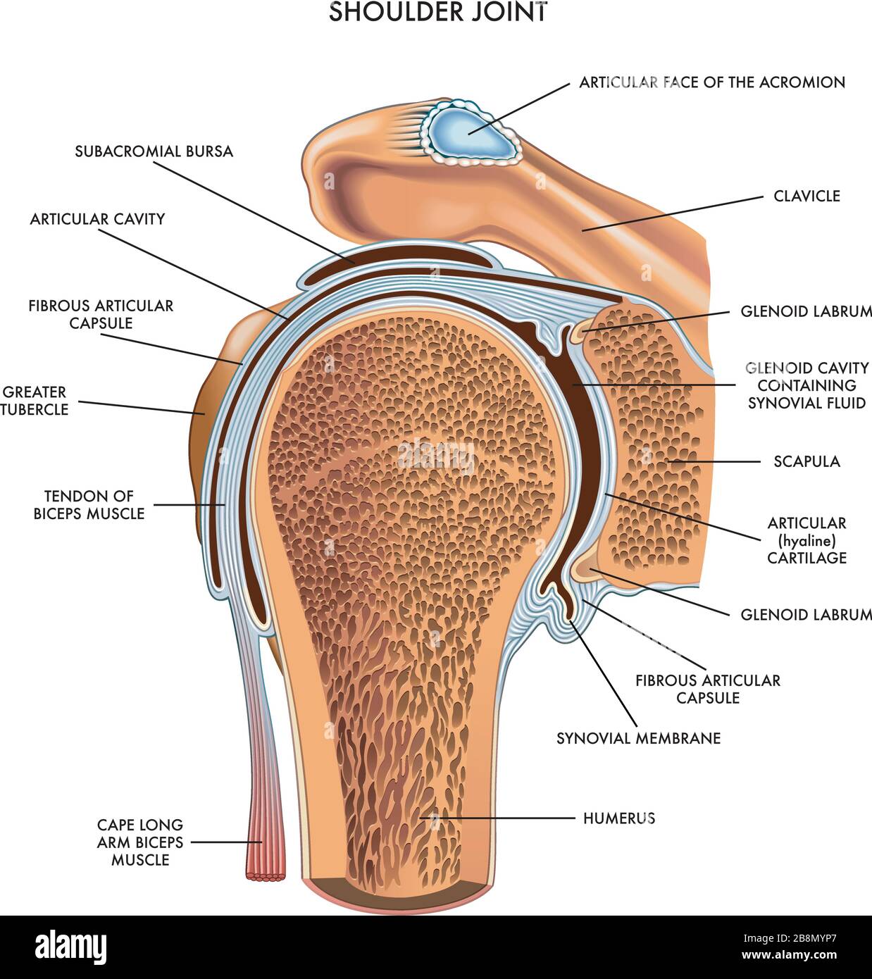
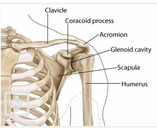
Shoulder pain that persists beyond a few days. The glenohumeral joint is a joint where the greater tubercle (humeral head at the top of the arm bone) meets the shoulder socket of the scapula, called the glenoid cavity or glenoid fossa. Muscles of the shoulder : The shoulder joint is protected superiorly by an arch, which is formed by the coracoid process of the scapula, the acromion process of the scapula and the clavicle. Four of them are found on the anterior aspect of the shoulder, whereas the rest are located on the shoulder's posterior aspect and in the back.
Shoulder Joint Anatomy High Resolution Stock Photography And Images Alamy from c8.alamy.com Learn faster with interactive shoulder quizzes, diagrams and worksheets. Numerous muscles help stabilize the three joints of. This diagram depicts muscle diagram of shoulder. The glenohumeral joint is a joint where the greater tubercle (humeral head at the top of the arm bone) meets the shoulder socket of the scapula, called the glenoid cavity or glenoid fossa. A second joint in the shoulder is the junction of the collar bone with the shoulder blade, called the. Four of them are found on the anterior aspect of the shoulder, whereas the rest are located on the shoulder's posterior aspect and in the back. Pronate your wrist so the palm of your hand faces down to the floor (as if you were trying to empty a glass of water). The shoulder has about eight muscles that attach to the scapula, humerus, and clavicle.
It is an extremely mobile joint, in which stability has been sacrificed for mobility.
The shoulder joint (glenohumeral joint) is a ball and socket joint between the scapula and the humerus.it is the major joint connecting the upper limb to the trunk. There are actually four joints that make up the shoulder. The supraspinatus is located on the upper part of the shoulder joint and is involved in abduction (arm raising). The glenohumeral joint, the acromioclavicular joint (a/c joint) and the sternoclavicular joint. The shoulder blade is called the scapula and the collarbone is called the clavicle. If you have new, worsening, or severe shoulder pain, you should seek medical attention. A diagram of an anatomic shoulder replacement—the plastic socket replaces the cup of the scapula (shoulder blade). The large bone in the upper arm is called the humerus. Medical labeled diagram closeup with muscle, transverse carpal ligament, median nerve, tendon sheath, flextor tendons and bones. Some signs that you should be seen by a doctor include: The treatment options are either replacement of just the head of the humerus bone (ball), or replacement of both the ball and the socket (glenoid). Shoulder muscles diagram / 3 : Sechrest, md narrates an animated tutorial on the basic anatomy of the shoulder.
The shoulder is a complex combination of bones and joints where many muscles act to provide the widest range of motion of any part of the body. What are common rotator cuff injuries? The shoulder blade is called the scapula and the collarbone is called the clavicle. Inability to carry objects or use your arm. This is the main muscle that lets you rotate and extend your shoulder.
Shoulder Joint Of Human Body Anatomy Infographic Diagram With Royalty Free Cliparts Vectors And Stock Illustration Image 87967058 from previews.123rf.com It is an extremely mobile joint, in which stability has been sacrificed for mobility. In this episode of eorthopodtv, orthopaedic surgeon randale c. Is the wear and tear of shoulder cartilage until bare bone is exposed. A metal ball component replaces the worn humeral head. The shoulder joint (glenohumeral joint) is a ball and socket joint between the scapula and the humerus.it is the major joint connecting the upper limb to the trunk. The scapula shoulder blade is a triangular shaped bone with 2 bony projections at the top right at your shoulder cuff. The glenohumeral joint is a joint where the greater tubercle (humeral head at the top of the arm bone) meets the shoulder socket of the scapula, called the glenoid cavity or glenoid fossa. Two joints are at the shoulder.
An injury that causes joint deformity.
Human anatomy diagrams show internal organs, cells, systems, conditions, symptoms and sickness information and/or tips for healthy living. Shoulder pain that occurs at night or while resting. Its main job is to assist with rotation of the arm away from the body. Injuries to the rotator cuff are common, but treatment is often successful. Muscles of the shoulder : The scapula shoulder blade is a triangular shaped bone with 2 bony projections at the top right at your shoulder cuff. The shoulder joint (glenohumeral joint) is a ball and socket joint between the scapula and the humerus.it is the major joint connecting the upper limb to the trunk. The muscles of the shoulder support and produce the movements of the shoulder girdle.they attach the appendicular skeleton of the upper limb to the axial skeleton of the trunk. Common rotator cuff injuries include rotator cuff tendonitis and rotator cuff strain, which is a partial or complete tear of the rotator cuff. This diagram depicts muscle diagram of shoulder. An injury that causes joint deformity. The shoulder is a complex combination of bones and joints where many muscles act to provide the widest range of motion of any part of the body. The human shoulder is made up of three bones:
This is the smallest rotator cuff muscle. Shoulder muscles diagram / 3 : What are common rotator cuff injuries? The muscles of the shoulder support and produce the movements of the shoulder girdle.they attach the appendicular skeleton of the upper limb to the axial skeleton of the trunk. Some signs that you should be seen by a doctor include:
Shoulder Anatomy Part 1 My Statement Blog 1306 from gtms1306.files.wordpress.com Bursitis (joint inflammation) cervical radiculopathy. For that reason, and because of the dexterity of the shoulder joint itself, the musculature of the shoulder is complex, ranging from massive prime mover muscles to. The shoulder joint (glenohumeral joint) is a ball and socket joint between the scapula and the humerus.it is the major joint connecting the upper limb to the trunk. A metal ball component replaces the worn humeral head. The shoulder is comprised of a ball (humerus) and socket (scapula), bones, ligaments, tendons and muscles that move the arms and connect them to the torso. Medical labeled diagram closeup with muscle, transverse carpal ligament, median nerve, tendon sheath, flextor tendons and bones. It is one of the most mobile joints in the human body, at the cost of joint stability. The shoulder joint can sometimes become narrowed and arthritic, and spurs can form on the undersurface.
The shoulder is one of the largest and most complex joints in the body.
The supraspinatus is located on the upper part of the shoulder joint and is involved in abduction (arm raising). Human anatomy diagrams show internal organs, cells, systems, conditions, symptoms and sickness information and/or tips for healthy living. Or a broken collarbone most often happens from falling and landing directly onto your shoulder, such as while bicycling, skiing, or. Smartdraw includes 1000s of professional healthcare and anatomy chart templates that you can modify and make your own. Inability to carry objects or use your arm. The scapula shoulder blade is a triangular shaped bone with 2 bony projections at the top right at your shoulder cuff. Medical labeled diagram closeup with muscle, transverse carpal ligament, median nerve, tendon sheath, flextor tendons and bones. Plus, exercises for training them. The shoulder joint is formed where the humerus (upper arm bone) fits into the scapula (shoulder blade), like a ball and. In this episode of eorthopodtv, orthopaedic surgeon randale c. The shoulder blade is called the scapula and the collarbone is called the clavicle. Avascular necrosis (death of bone tissue due to limited blood flow) brachial plexus injury. The shoulder is a complex combination of bones and joints where many muscles act to provide the widest range of motion of any part of the body.


Post a Comment